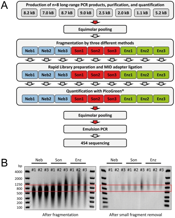
Figure 1. Workflow for fragmentation and NGS sequencing of long-range PCR fragments.
(A) Graphical illustration of the entire workflow. The red arrows depict a measuring and DNA-quantification step. (B) Analysis of fragment lengths by PAGE before (left panel) and after (right panel) removal of small fragments <500 bp with AMPure™ columns. The red boxes depict the desired size range between 600 and 1,000 bp. Neb, nebulization; Son, sonication; Enz, enzymatic fragmentation.

