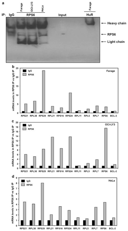
Figure 2.
RPS6 associates with multiple 5′ TOP messages. (a) Representative immunoprecipitation assays performed as described (Materials and methods) but followed by western blot analysis to assess the abundance of RPS6 protein in the IP material, isotype IgG and non-RPS6-binding protein HuR were included as controls. (b–d) Cytoplasmic lysates from Farage, OCI-LY3 and HeLa cell lines were used for immunoprecipitation assays, using anti-RPS6 antibody. RNA was isolated and reverse transcription followed by qPCR was performed to measure abundance of RNA. Graph represents the mean from three independent assays.

