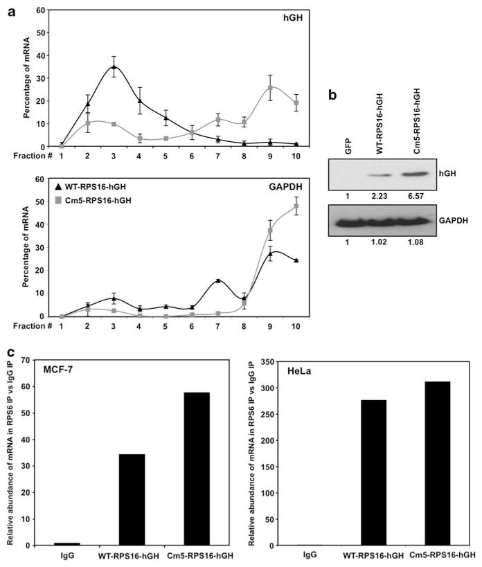
Figure 3.
RPS6 associates with both wild-type and mutant 5′ TOP messages. (a) Quantitative PCR analysis of the distribution on polysomes of hGH reporter mRNA in HeLa cells transfected with either WT-RPS16-hGH or Cm5-RPS16-hGH plasmids (b) Representative western blot analysis of HeLa cells transfected with either control green florescent protein (GFP), WT-RPS16-hGH or Cm5-RPS16-hGH. A volume of 10 μg of total protein lysates was loaded and the abundance of hGH and glyceraldehyde 3-phosphate dehydrogenase (GAPDH) was assessed. (c) Cytoplasmic lysates from MCF-7 and HeLa cell lines were used for immunoprecipitation assays, using anti-RPS6 antibody. RNA was isolated and reverse transcription followed by qPCR was performed to measure abundance of RNA. Graph represents the mean from three independent assays.

