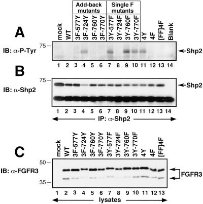
Figure 5.
Tyrosine phosphorylation of the FGF-responsive effector protein Shp2. (A) Detection of tyrosine-phosphorylated Shp2 in FGFR3-expressing cells. (B) The same membrane in A was stripped and reprobed with Shp2 antiserum, to confirm approximately equal recovery of Shp2 protein in each sample. (C) Whole-cell lysates were analyzed by immunoblotting with FGFR3 antiserum to confirm equivalent expression levels of each construct.

