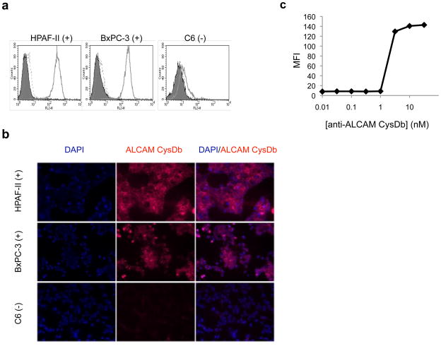
Figure 3.
Functional characterization of anti-ALCAM CysDb. a, Evaluation of binding specificity by flow cytometry. Left, HPAF-II; middle, BxPC-3; right, C6. Filled peak, cells only. Solid line, cells incubated with anti-ALCAM CysDb, followed by mouse anti-Penta-His antibody and PE-conjugated goat anti-mouse antibody. Dashed line, cells incubated with only mouse anti-Penta-His antibody and PE-conjugated goat anti-mouse antibody. b, Evaluation of binding to cultured cells by immunofluorescence. Top panels, HPAF-II; middle panels, BxPC-3; bottom panels, C6. Left column, DAPI staining. Middle column, Alexa Fluor 647-anti-ALCAM CysDb staining. Right column, overlay. c, Determination of KD by flow cytometry.

