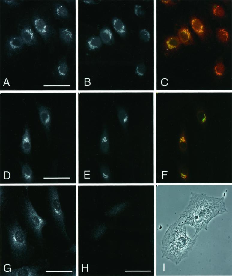
Figure 2.
Immunolocalization of PLD1 in rat NRK cells. Cells were stained with rabbit antibodies directed against PLD1 peptides P1–P4 (A and D). These cells were costained with either mAbs 53FC3 directed against mannosidase II (B) or GM130 (E). C and F show the overlaping regions between PLD1 and the Golgi marker proteins. (G) Cells were also stained with an independently generated rabbit antibody to the C-terminal of PLD1 (MATERIALS AND METHODS). (H) Preincubation of the P1–P4 antibody with 1.3 μg of antigenic peptide before immunolocalization; (I) corresponding phase-contrast image. Images are from projected Z-series. Bar, 10 μm.

