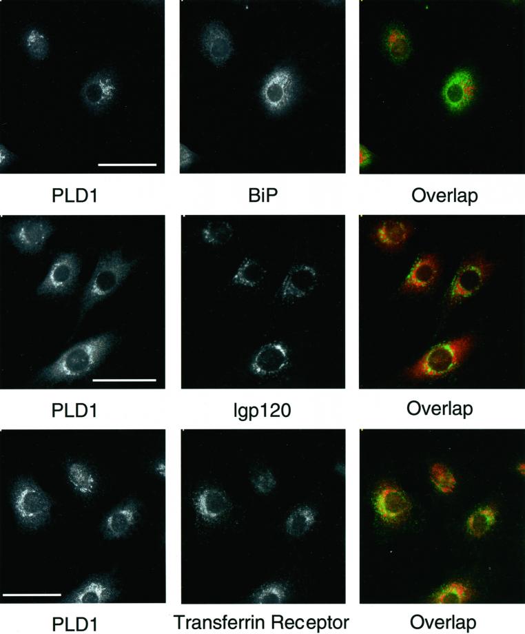
Figure 3.
PLD1 localization with different organelles in rat NRK cells. Cells were prepared for immunofluorescence microscopy and incubated with rabbit anti-P1–P4 antibodies to PLD1 and costained with mAbs to the ER protein BiP, with the late endosome/lysosome marker lgp 120, or with a marker for the plasma membrane and early endosomes, transferrin receptor. Note the partial overlap between PLD1 with lgp120 and transferrin receptor but only minimal colocalization with BiP. Bar, 10 μm.

