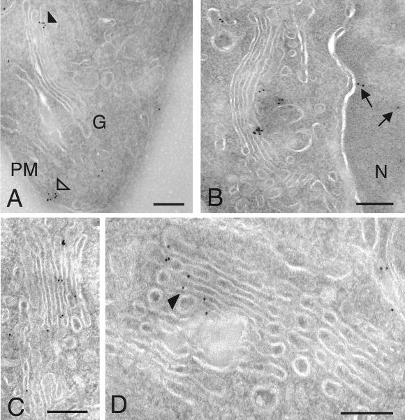
Figure 4.
Localization of PLD1 by cryo-immunoelectron microscopy. GH3 cells were prepared for cryo-immunoelectron microscopy (MATERIALS AND METHODS). Sections were labeled with polyclonal antibodies directed against PLD1 (P1–P4) followed by secondary antibodies conjugated to 10-nm gold particles. Staining revealed PLD1 localization to the plasma membrane (A, PM, open arrowhead) and Golgi apparatus (G, indicated by filled arrowheads in A and D), as well as throughout the Golgi cisternae (B–D) and nuclei (B, N, arrows). Bars, 0.2 μm (B–D); 0.5 μm (A).

