Figure 5.
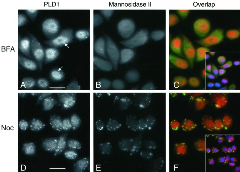
Brefeldin A and nocodazole alter the localization of Golgi-associated PLD1. GH3 cells were incubated with 5 μg BFA/ml for 40 min (A–C) or with 10 μM nocodazole for 4 h at 37°C (D–F). After incubation, the cells were prepared for microscopy using the rabbit PLD1 antibody (P1–P4) or the 53FC3 monoclonal antibody to mannosidase II. PLD1 and mannosidase II were visualized using appropriate anti-rabbit and anti-mouse IgG antibodies (Figures 1 and 2). (C and F) Inset, nuclear staining with Hoechst 33258 dye. Note overlap between cell nuclei and PLD1. Bar, 10 μm. (A) Arrow indicates PLD1 enrichment in cell nuclei.

