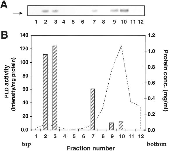
Figure 6.
PLD1 enzyme activity cofractionates with the Golgi apparatus in endocrine cells. GH3 cells were incubated with 10 μCi/ml 3H-oleic acid for 24 h to radiolabel phospholipids, after which the cells were homogenized. The homogenate was fractionated on a floatation gradient designed to separate the Golgi apparatus from total microsomes (MATERIALS AND METHODS). (A) An aliquot of each gradient fraction was assayed for ARF-1–stimulated PLD activity in the presence of 0.3% 1-BtOH, and the products were analyzed by TLC followed by fluorography. The arrow indicates the mobility of PtdBtOH. (B) The TLC plate was scanned using a densitometer, and the band intensities corresponding to PtdBtOH were quantitated using the Image Quant program (Siddhanta et al., 2000). An aliquot of each gradient fraction was also assayed for protein concentration. Hatched bars, PLD activity expressed as pixel intensity per milligram protein. Dashed line, distribution of total protein (mg/ml). TGN38 is concentrated in fractions 2 and 3 (Austin and Shields, 1996).

