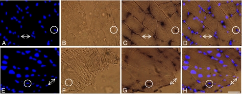
Fig. 1.
Localization of hsp70 mRNA in cross (a, b, c, d) and longitudinal sections (e, f, g, h) of the fast, white portion of the vastus lateralis, 1 h post-exposure to the positive control stimulus, 42°C heat stress, (×40 magnification). a, e Blue fluorescence of nuclear counterstain, DAPI. b, f A negative control for the in situ hybridization, sense probe, did not produce any non-specific signal in all samples examined. c, g In situ hybridization of hsp70 mRNA with antisense probe produced a concentrated dark and punctate signal near the periphery of the myofiber, and a diffuse signal throughout the cytoplasm. d, h Merged images of a and c or e and g demonstrated that all punctate hsp70 mRNA signal were associated with the nuclear counterstain DAPI (circles). Differential punctate hsp70 mRNA signal was found among the nuclei of individual myofibers; also consistent for all treatment groups (arrows). Bar = 50 μm

