Fig. 2.
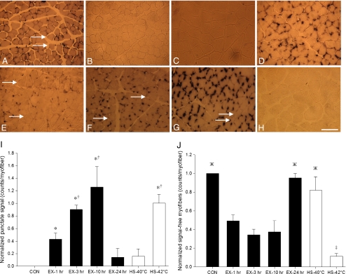
Temporal localization and temperature-related induction of hsp70 mRNA in situ hybridization signal in skeletal myofiber cross sections (×16 magnification). a Mixed-muscle, plantaris, taken from sedentary control animals, displayed constitutive hsp70 mRNA signal that was concentrated in a punctate fashion. b–h Fast-muscle, white vastus. b Sedentary control displayed no detectable hsp70 mRNA signal. e, f, g, and h follow the temporal localization of hsp70 mRNA signal at 1, 3, 10, and 24 h after a single bout of intense exercise, respectively. hsp70 mRNA signal was observed to be concentrated in a punctate fashion with a concomitant tend in rising counts of punctate signal per myofiber over time such that: b < e <f < g, before falling back to resting levels 24 h post-exercise treatment (h). Although not quantified, the intensity of punctate signal also appeared to rise up to 10 h post-exercise. c and d display hsp70 mRNA signal 1 h post-exposure to 40°C and 42°C heat stress, respectively; 40°C heat stress treatment (c) represents a temperature similar to that experienced during exercise, but it was observed to promote less punctate hsp70 mRNA signal than 1 h post-exercise (e) and 42°C heat stress (d). i The histogram illustrates the counts of punctate hsp70 mRNA signal per myofiber for the various treatment groups. *Significantly greater than CON, EX-24 h, and HS-40°C. †Significantly greater than EX-1 h. j The histograms illustrates the counts of hsp70 mRNA signal-free myofibers (arrows). ‡P < 0.05, significantly less than all other groups. ЖP < 0.05, significantly greater than EX-1 h, EX-3 h, and EX-10 h. All histogram values are normalized mean (n = 5) ± SE and considered significant at P < 0.05. In histograms, black represents control and exercise treatments, while white represents heat stress treatments. Bar = 125 μm

