
Figure 2. Lin28A and Lin28B are differentially localized within the cell.
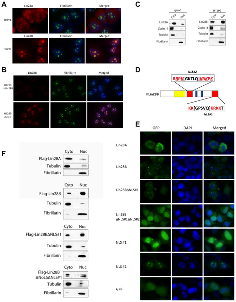
(A) Immunofluorescence detection of endogenous Lin28A in Igrov1 and Lin28B in H1299 cell lines. Fibrillarin, a known nucleolar protein, was used as a positive control. (B) Immunofluorescence analysis of control- and Lin28B-knockdown H1299 cell lines. (C) Biochemical fractionation of Igrov1 and H1299 cell lines. Endogenous Lin28A, Lin28B, and Zcchc11 in each fraction were detected by western blot. Fibrillarin was used as a nuclear marker; Tubulin was used as a cytoplasmic marker. (D) Schematic of nuclear localization signals (NLS) in the Lin28B protein. An Arginine as well as several Lysines that were replaced by Glycines are underlined and italicized (E) Localization of GFP-Lin28 fusion proteins in Hela cells. (F) Fractionation of Flag-Lin28 proteins, exogenously expressed in Hela cells. Proteins were detected by Flag western blot.