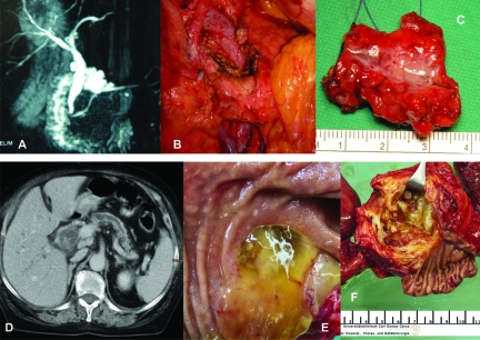
Figure 5.
Clinical examples of IPMN. (A–C): BD-IPMN. (A): MRCP; (B): Intraoperative situs after enucleation; (C): Surgical specimen with BD-IPMN. (D–F): MD-IPMN. (D): CT with cystic head tumor and enlarged main pancreatic duct; (E): Mucin extrusion from a widely patent ampulla of Vater; (F): Surgical specimen with MD-IPMN.
Abbreviations: BD-IPMN, branch duct IPMN; CT, computed tomography; IPMN, intraductal papillary mucinous neoplasm; MD-IPMN, main duct IPMN; MRCP, magnetic resonance cholangiopancreatography.

