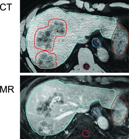
Figure 4.
Multimodality contrast imaging to aid in liver metastasis delineation. Both computed tomography (CT) and magnetic resonance (MR) imaging are done in an exhale breathhold to minimize differences in the liver shape and aid in fusion. The liver contour from the planning CT (in blue) is overlaid on the MR image, demonstrating excellent liver-to-liver registration.

