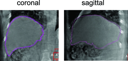
Figure 6.
Example of verification imaging (kilovoltage cone beam computed tomography [CT]) obtained in the radiation therapy treatment room, used to position a patient with liver cancer prior to conformal radiation therapy delivery. The pink contour, representing where the liver should be positioned (obtained from the planning CT), is overlaid on the verification cone beam CT image.

