Figure 5.
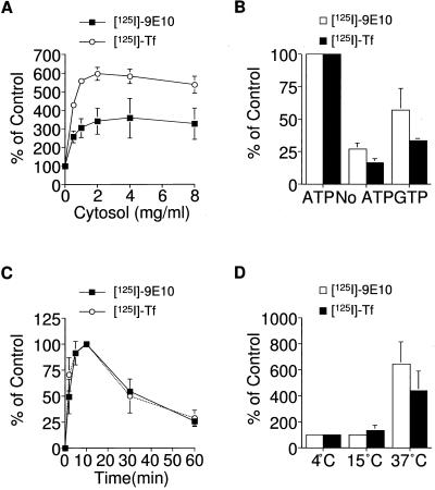
Characterization of the cell-free assay for ESV formation. (A) Cytosol concentration dependence of ESV biogenesis. Membranes (1 mg/ml) labeled with either 125I-9E10 or 125I-Tf were incubated with varying concentrations of rat brain cytosol in the presence of an ATP-regenerating system. Values are normalized relative to the minus-cytosol control. (B) Nucleotide dependence of ESV biogenesis. Budding from labeled membranes was carried out in the presence of cytosol with ATP, GTP, or no added nucleotide. Values were normalized relative to the amount of budding observed in the presence of 1 mM ATP. (C) Time course of ESV budding from labeled membranes at 37°C. Values were normalized relative to the maximal amount of ESV formation observed at 10 min. (D) Temperature dependence of ESV biogenesis. Budding from labeled membranes at either 0, 15, or 37°C. Values were normalized relative to the amount of budding observed at 0°C. The amount of specific radioactivity associated with de novo ESVs on glycerol gradients was determined for each reaction condition.

