Figure 7.
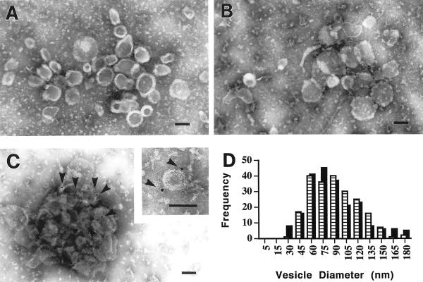
Negative stain electron micrographs of ESVs isolated from intact cells and generated in vitro. (A) ESVs isolated from intact CHO/GLUT4myc cells. (B) Vesicles generated in vitro from CHO/GLUT4myc cell membranes. (C) In vitro generated vesicles immunogold labeled with anti-GLUT4 polyclonal antibody R820. The inset shows a magnified image of a single in vitro generated vesicle carrying two gold particles (arrowheads). (D) Histogram showing the size distributions of in vivo (hatched bars) and in vitro (solid bars) generated vesicles. Both types of vesicles show a similar size distribution with a median external diameter of ∼70–90 nm. Negative stain, 2% ammonium molybdate. Bars, 100 nm.

