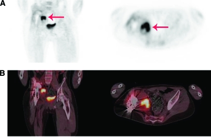
Figure 3.
An 18-year-old man with a megaprosthesis of the right hip after resection of Ewing's sarcoma was admitted with a painful hip 11 months after insertion of the prosthesis. Pseudomonas aeruginosa, Enterococcus faecium, and S. aureus were cultured from several tissue biopsies. (A): PET. (B): Fused PET-CT. Left panels show coronal view, right panels show transverse view. Intense FDG uptake adjacent to the prosthesis (red arrow) caused by infection is seen.
Abbreviations: CT, computed tomography; FDG, fluorodeoxyglucose; PET, positron emission tomography.

