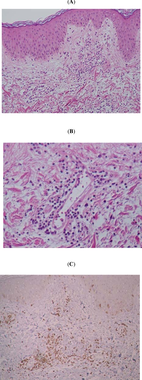
Fig. (3).
Histological and immunohistological characteristics of the pricked skin sites. Histology revealed interstitial edema and intense lympho-histiocytic infiltration (A, x40), but not a typical feature of vasculitis (B, x200), in the upper dermis. These inflammatory infiltrates are mainly composed of CD4+ T cells (73.4%) and to lesser with CD8+ T cells (12%) and CD68+ cells (38%) (C). No other cell sources, such as CD20+ or CD56+ cells were found.

