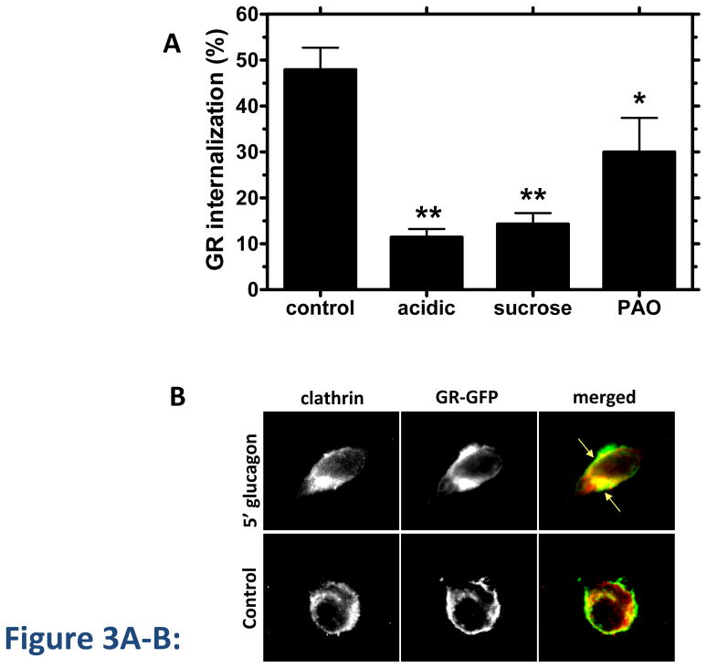
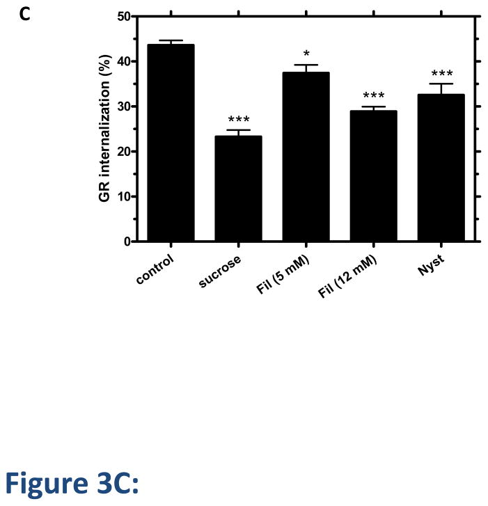
Figure 3. GR internalization involves clathrin and caveolae.
A, HEK-GR cells were serum-starved for 1 h and treated with inhibitors of clathrin-mediated endocytosis: acidic medium (pH 5.5), hypertonic medium (0.45 M sucrose), or 20 μM phenylarsine oxide (PAO) for 30 min, followed by 30 minute incubation with 100 nM glucagon. Control cells were incubated with glucagon alone. GR internalization was determined by binding (Method B). Data represent the mean ± s.e.m. of 3 independent experiments.*, p < 0.05 vs. control, **, p < 0.01 vs. control. B, HEK-293 transfected with GR-GFP were serum-starved for 1 h and incubated with or without (control) 100 nM glucagon for 5 min, fixed, permeabilized and immunostained for clathrin using an anti-clathrin heavy chain antibody. Each fluorescence image is a deconvolved projection of 5–6 planes acquired using a 60 × objective and is representative of 3 experiments. Arrows indicate colocalization of clathrin and GR-GFP. C, HEK-GR cells were pre-treated for 30 min with filipin III (Fil., 5 μM or 12 μM), 0.45 M sucrose or 5 μg/mL nystatin (Nyst). GR internalization after 100 nM glucagon incubation for 30 min was measured by binding (Method A). The data represent the mean ± s.e.m. of 4–5 independent experiments. *, p < 0.05, **, p < 0.01; ***, p < 0.001 vs. control.


