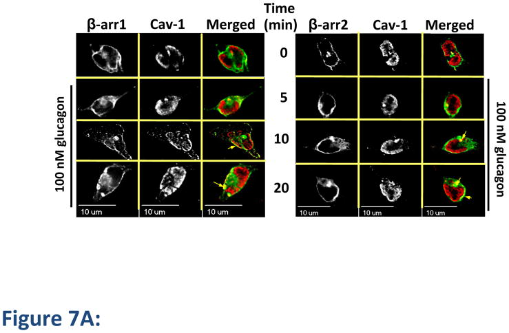
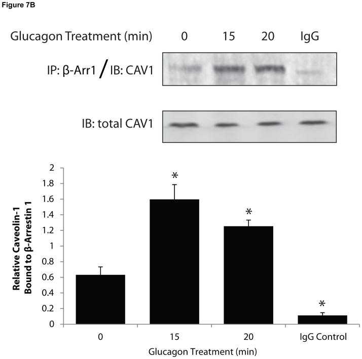
Figure 7. Caveolin-1 colocalizes with β-arrestins.
HEK-FLAG-GR cells were transfected with either β-arrestin1-GFP (β-arr1, green) or β-arrestin2-GFP (β-arr2, green). The cells were starved for 1 h prior to treatment with glucagon. A, After treatment, the cells were fixed, permeabilized and immunostained with anti-Cav-1 antibody (cav-1, red). Each fluorescence image is a deconvolved projection of 5–6 planes acquired using a 60× objective. Representative images from 3 independent experiments are shown. Arrows indicate colocalization of caveolin-1 and β-arrestins. B, After treatment, cells were lysed and β-Arr1 was immunoprecipitated with an anti-GFP antibody. Immunoprecipitated lysates were immunoblotted for caveolin-1 (CAV1). In parallel experiments, an IgG control was used for immunoprecipitation followed by immunoblotting for CAV1. Top panel is a representative blot; bottom panel represents the mean + S.E.M. of 3 independent experiments. *Significantly different from the 0 time point (P < 0.05).


