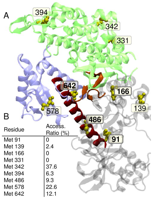
Fig. 1.
A. Location of native methionines in Dictyostelium myosin II (1FMV). Lower 50 kDa domain (blue); upper 50 kDa domain (green), relay helix (red); nucleotide binding pocket (orange); methionines (yellow spheres). Conserved Met residues are shown bold. B. Accessible surface area calculated from the crystal structure 1FMV and reported as a ratio of accessible side-chain surface area to the value calculated for a random coil (53). VMD (Visual Molecular Dynamics) was used to create renderings of molecular structures (54).

