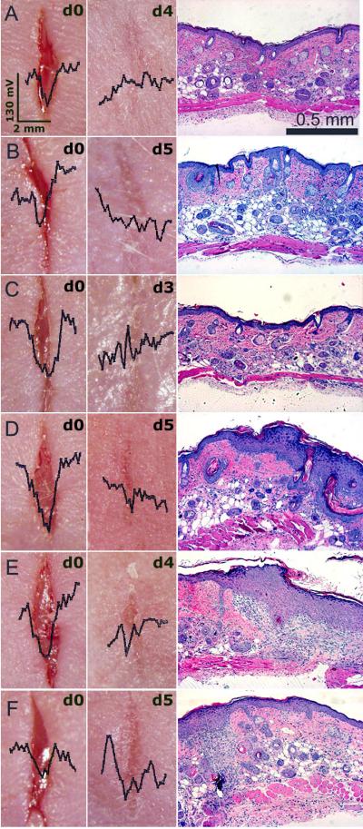
Figure 6.
Dermacorder® scans of 6 wounds just after creating them and 3-5 days later. Histological section on the right of each wound was prepared from the excised skin wound pictured to the left of it. A-C: Wounds that formed a compact epidermal layer exhibited little to no electric field. D-F: Wounds that were more slowly healing and exhibited diffuse epidermal layers still exhibited an electric field 3-5 days after wounding. The scale bar on the wound photo in “A” applies to all of the wound photos and the scale bar in the histological section in A applies to all sections beneath it.

