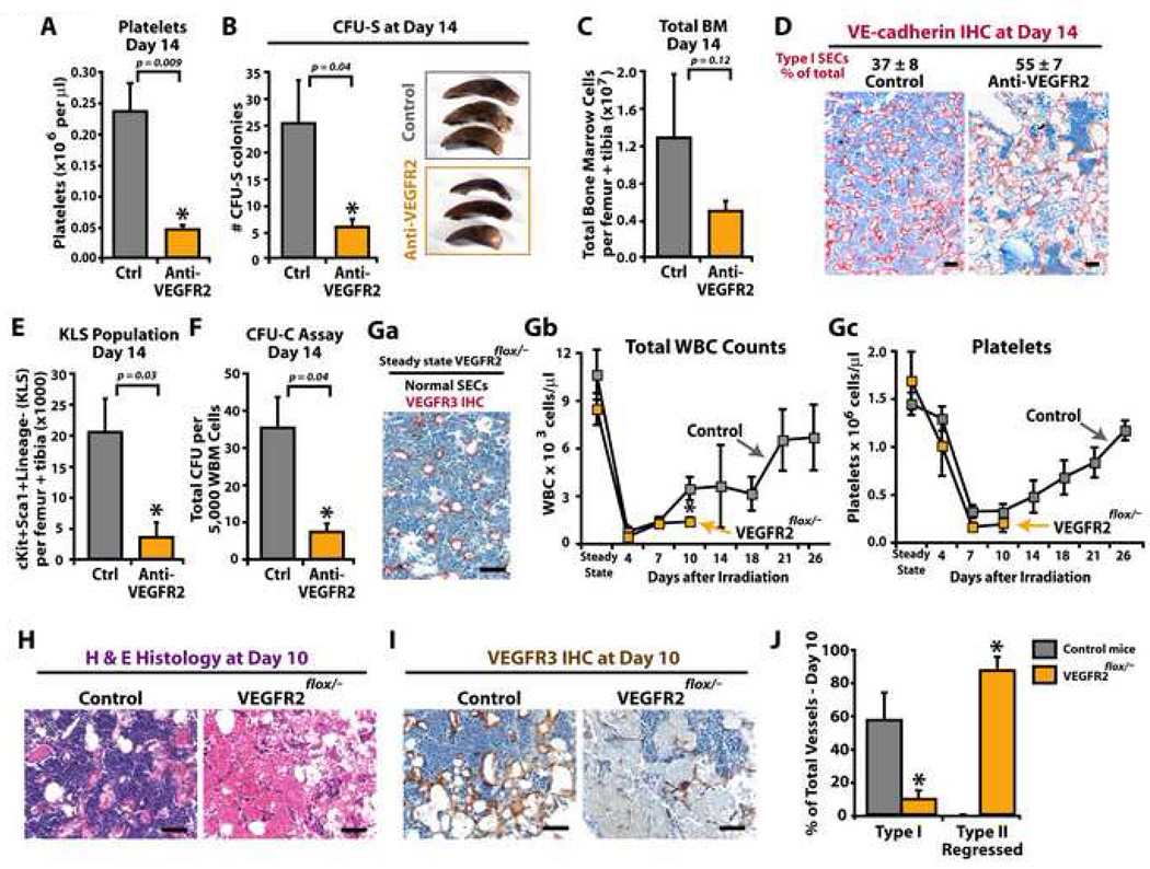
Figure 5. Impaired regeneration of SECs after 650 rad irradiation results in failure of hematopoietic reconstitution.
The consequence of 650 rad in the reconstitution of hematopoiesis was evaluated in WT (A–F) and VEGFR2 conditional knockdown (VEGFR2flox/−) mice (G–J). A–F) WT mice were treated with 650 rad with and without neutralizing mAb to VEGFR2 (DC101, 800 µg/dose thrice weekly). At day 14, PB, spleens and BM were harvested for evaluation of (A) platelets, (B) CFU-spleen forming units (CFU-S), (C) total BM mononuclear cells, and (D) IHC quantification of Type I VE-cadherin+ vessels. E–F) BM mononuclear cells were evaluated for KLS population (E), and colony forming units (CFU-C) (F). G–J) Groups of ROSA-CreERT2VEGFR2e3loxP/− mice and control WT and ROSA-CreERT2VEGFR2e3loxP/+ mice were treated with tamoxifen and allowed to recover. Mice were given 650 rad, and hematopoietic recovery and vessel integrity were monitored. Ga) Steady state untreated femurs from VEGFR2flox/− mice were immunostained with anti-VEGFR3 Abs and vascular integrity of SECs (normal versus Type I/II vessels) was assessed. Virtually all vessels in VEGFR2flox/− mice prior to irradiation are normal SECs. Gb-c) WBC (Gb) and platelets (Gc) were counted at designated time points. At steady state conditions after tamoxifen treatment, there is no difference between the WBC and platelets counts as compared to non irradiated controls. H–J) At day 10, femurs were harvested and SEC status was evaluated using H&E (H) and anti-VEGFR3 IHC (I). J) VEGFR3 stained BM sections were assessed for Type I and Type II SECs. Although the control animals consistently show signs of SEC recovery, VEGFR2 knockdown animals have a profound persistence in Type II regressed SECs. Micron bar = 50 µm.

