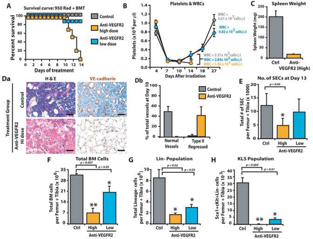
Figure 6. Engraftment and replenishment of HSPCs require VEGFR2 dependent regeneration of regressed SECs.
WT mice were treated with 950 rad + BMT in combination with mAb to VEGFR2 (DC101) at high dose (800 µg/dose) or low dose (400 µg/dose), or control IgG, thrice weekly. At day 13, PB, BM and spleen were processed for analyses. A) Survival plot for mice given 950 rad + BMT in combination with anti-VEGFR2 mAb treatment (low and high, blue and orange, respectively) vs. control (grey) demonstrating that 100% of mice treated with high dose mAb succumbed to hematopoietic failure even after BMT (n=10, P<0.001). B) Platelet and WBC counts are decreased in mice treated with high dose (n=6, P<0.006 as compared to low dose, orange circles), but not low dose, mAb to VEGFR2 (blue circles) or IgG control (gray circles). C) As compared to IgG control mice, irradiated 950 rad + BMT treated mice injected with high dose, but not low dose, anti-VEGFR2 mAb showed a marked decrease in splenic mass (n=6, P<0.001). D) IHC for BM vessels using VE-cadherin (Da) reveals a predominance of Type II VE-cadherin+ regressed BM vessels (Db) (n=5, P<0.04). E) Total number of SECs was quantified by polyvariate flow cytometry. Calculating the total number of VEGFR3+VEGR2+VE-cadherin+CD31+CD45−CD11b−Sca1− vessels demonstrated a reduction in SECs in high dose mAb treated mice vs. control (n=3 P<0.04). F–H) Treatment of 950 rad + BMT irradiated mice with high dose or low dose mAb to VEGFR2, but not control IgG, resulted in a statistically significant decrease in total BM mononuclear cells (F, P<0.007 for high dose, P<0.05 for low dose), decrease in Lin− cells (G, P<0.02 for high dose, P<0.03 for low) and virtually absent KLS (H, P<0.004) in high dose and diminished KLS (P<0.01) in low dose treated mice. Micron bar = 50 µm.

