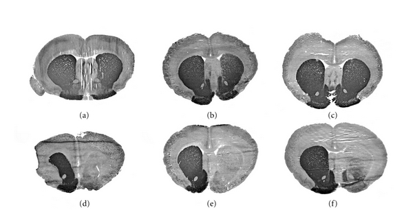
Figure 4.

Levels of TH in brain following TH-gene therapy in the 6-OHDA Parkinson's disease model. The TH immunocytochemistry was performed in rat brains removed 72 hours after a single intravenous injection of 10 μg per rat of clone 951 plasmid DNA encapsulated in THL targeted with either the TfRMAb (a, b, and c) or with the mouse IgG2a isotype control (d, e, and f). Coronal sections are shown for 3 different rats from each of the two treatment groups. The 6-hydroxydopamine was injected in the medial forebrain bundle of the right hemisphere, which corresponds to right side of the figure. Sections are not counterstained. The animals that received the TH gene therapy had a normalization of the brain TH levels as compared to the animals administered the nontargeted THLs, which showed complete lost of immunoreactive TH in the same region. From [30].
