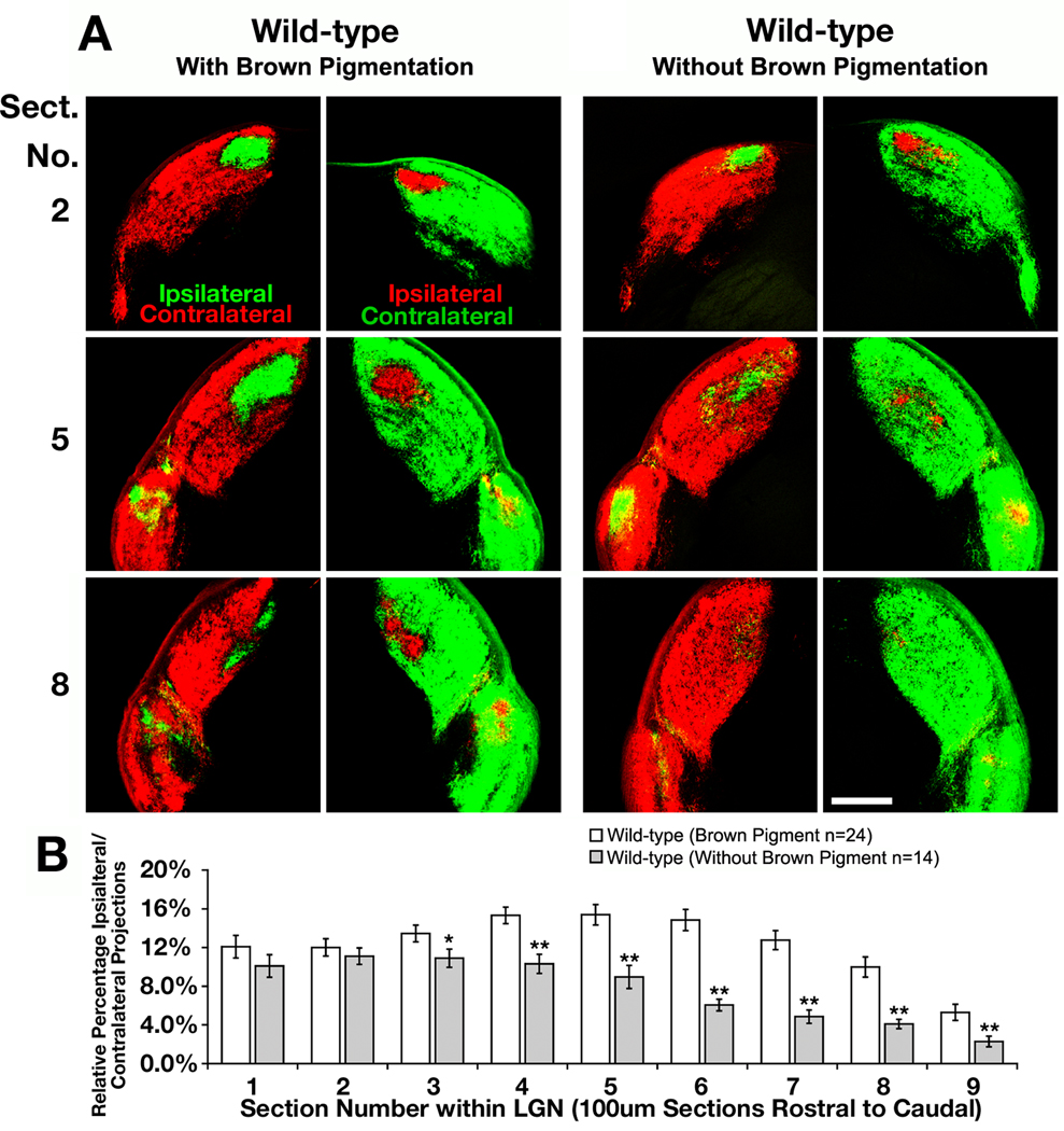
Fig. 3.
Brown pigmented mice possess a larger percentage of ipsilateral projections to the lateral geniculate nucleus than mice lacking brown pigmentation. (A) Serial sections through the lateral geniculate nucleus of a wild-type mouse with brown pigmentation and another without brown pigmentation. The retinal ganglion cells were labeled with cholera toxin subunit-B-555 (CTB-555, red) anterograde dye in right eye and CTB-488 (green) in the left eye to compare changes in the amount of ipsilaterally projecting RGC axons. Scale bar = 250 µm. (B) Quantitative analysis showing the differences in relative percentage of ipsilaterally projecting RGC axons throughout the entire lateral geniculate nucleus (LGN) of each genotype (* indicates p<0.05, ** indicates p<0.01).

