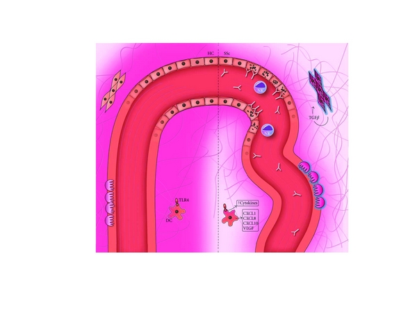
Figure 1.

Hypoxia in the pathogenesis of systemic sclerosis. The left side illustrates the normal situation, with a healthy blood vessel delivering oxygen to the surrounding tissue. The right side represents the situation in SSc, where the diseased vessel and overwhelming deposition of collagen fibers prevent the oxygen from reaching the periphery. In the vessel, endothelial apoptosis as a result of antibody-dependent cytotoxicity is visible. Surrounding the ECs, activated pericytes are responsible for the deposition of collagen. The resulting hypoxia leads to a higher cyto- and chemokine production by DCs, in part triggered by TLR stimulation, and to a continuing loop of TGFβ production, collagen synthesis, and myofibroblast differentiation of fibroblasts. This in turn leads to a hindered dispersion of oxygen, keeping the vicious circle going on.
