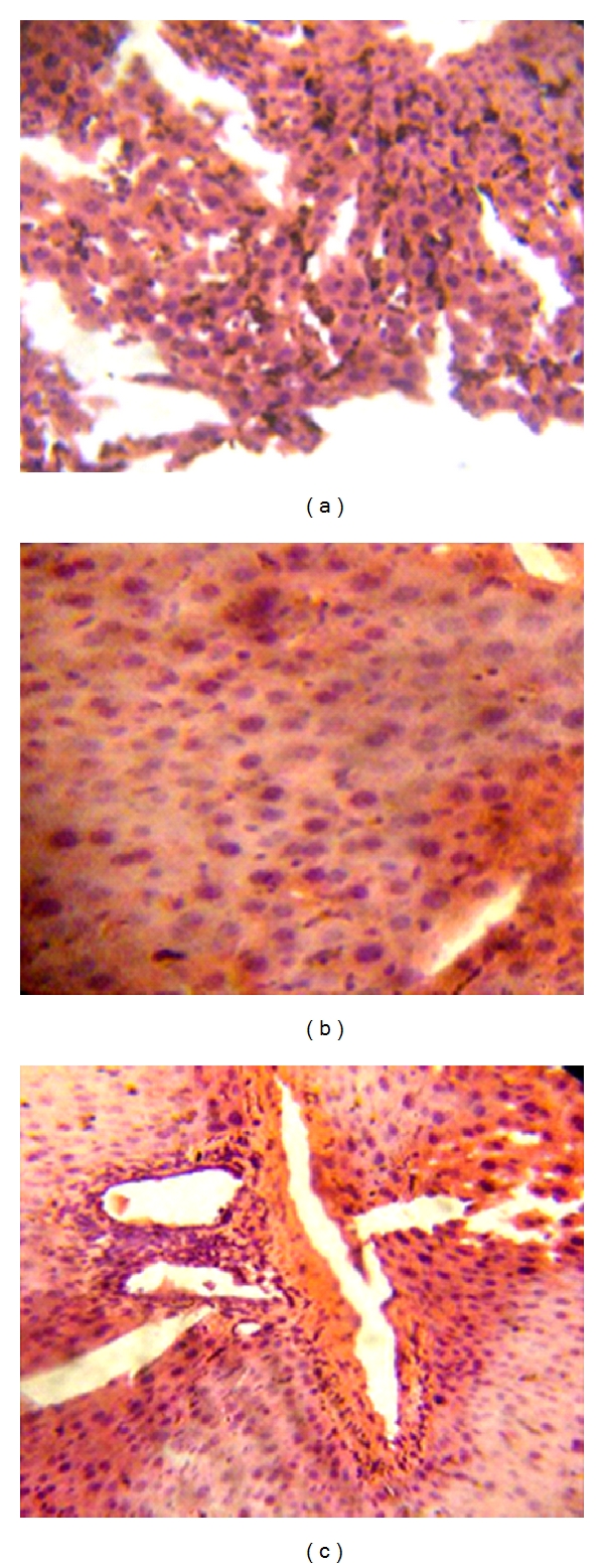
Figure 3.

(a) Liver photomicrograph of Plasmodium berghei-infected mice showing dilated hepatic sinusoids congested with hypertrophied Küpffer's cells-laden malaria pigment and parasitized red blood cells (Haematoxylin and Eosin stain, ×200), (b) Liver photomicrograph of Plamodium berghei-infected mice treated with 300 mg/kg dose of Ficus platyphylla Del. Micrograph shows progressive clearance of Küpffer's cells-laden malaria pigment (Haematoxylin and Eosin stain, ×200), (c) Liver photomicrograph of naive experimental control mice showing normal lobular architecture of the liver (Haematoxylin and Eosin stain, ×200).
