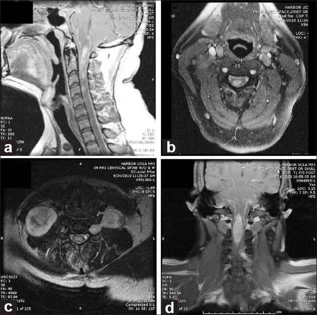
Figure 1.

MRI of the neck with gadolineum. a) Sagittal T1 image illustrating an intramedullary enhancing mass from the cervicomedullary junction to C4. Leptomeningeal enhancement is also present. b) Axial T1 image illustrating the intramedullary mass and leptomeningeal enhancement. c) Axial image of cervical spine illustrating a dumbbell mass extending through C5-C6 neural foramen and paraspinal mass. d) Coronal T1 image illustrating intramedullary mass
