Figure 3.

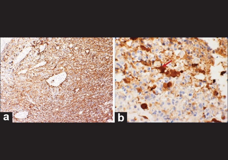
Immunohistomchemical staining. a) GFAP positive b) Synapthophysin positive, illustrating positivity around a binucleated cell (red arrow)

Immunohistomchemical staining. a) GFAP positive b) Synapthophysin positive, illustrating positivity around a binucleated cell (red arrow)