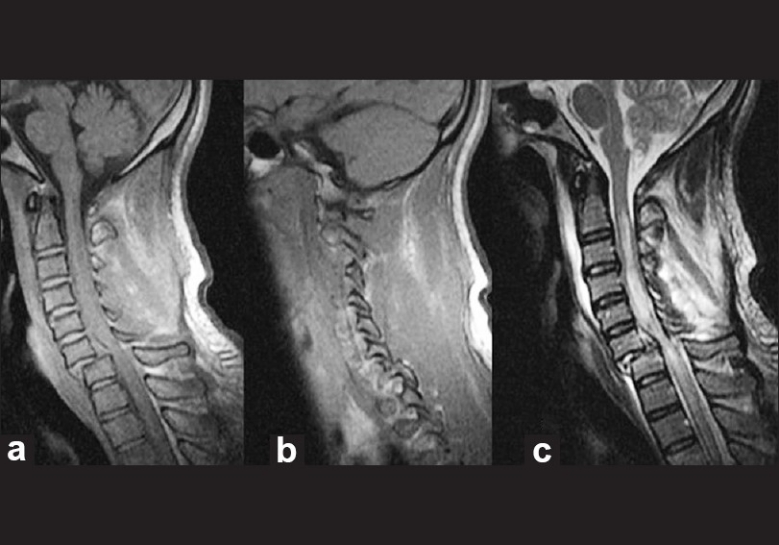
Figure 1.

T1- and T2-weighted sagittal MRI scans showing the traumatic spondylolisthesis at C6/C7 (a-c). The image b demonstrates the facetal dislocation. The image on the right (c) shows the related spinal cord injury and a posterior disc fragment. This precludes spinal traction before reduction as the disc may be pushed back into the spinal cord causing more injury
