
Figure 5.
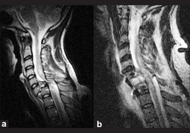
MRI scan of the patient (T2 sagittal images) showing the level of injury and dislocation, C6/C7 with spinal cord transection. Pre-operative image (a). The disc prolapse behind the vertebral body is significant. The post-operative image (b) is also shown following anterior approach, discectomy, and fixation with a hip graft and plate