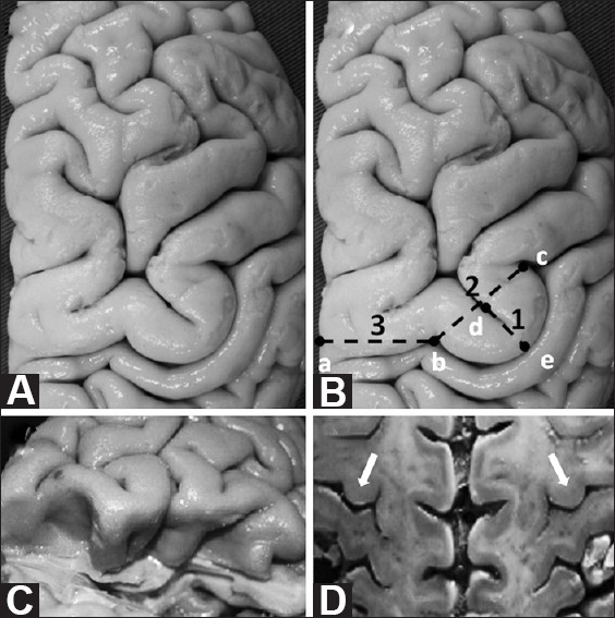
Figure 1.

(A and B) Superior view of the Omega sign in a right brain. (B) Measurements taken in this study are highlighted in this diagram, based on the specimen shown in (A): 1(d-e) Omega height; 2(b-c) Omega width; 3(a-b) distance between the medial edge of the hemisphere and the medial limit of the Omega. (C) Post-central gyrus excised to expose pre-central gyrus. Note that the Omega shape of the central sulcus remains deep within the sulcus. (D) An axial section through both hemispheres resembles an MRI axial section. Note that both Omegas are at the same anterior posterior level, an anatomical fact that enables the use of the contralateral Omega sign for preoperative planning when the ipsilateral anatomy is distorted owing to a disease process
