
Figure 5.
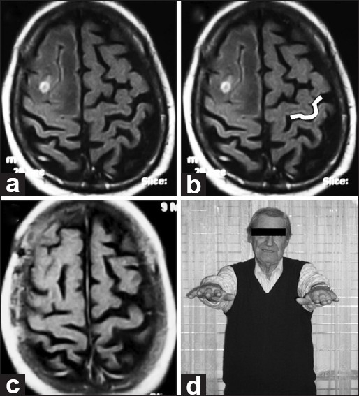
(a and b) Pre-op axial MRI. Imaging reveals a small lesion with perilesional edema involving the right middle frontal gyrus posteriorly and distorting normal right-brain anatomy. On the opposite side, the Omega shape is highlighted in white. (c) Post-op axial MRI. Imaging demonstrates appropriate excision. Histologically, a lung cancer metastasis was found. (d) Follow-up. No motor deficits seen at neurological examination