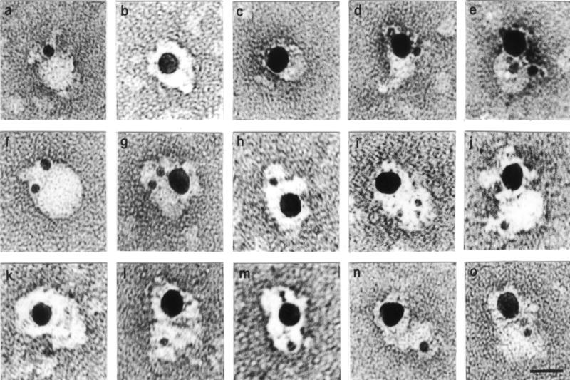
Figure 11.
EMTCs morphology and immunocytochemistry at the electron microscope level. EMTCs present in fraction 5 (a, c, d, and e) of glycerol gradient have been immunolabeled as described in MATERIALS AND METHODS with 5 nm gold-conjugated anti-myc mAb and/or 15 nm gold-conjugated anti-caveolin pAb and with 15 nm gold-conjugated anti-NSF pAb (b). Cytosolic EMTCs present in fraction 10 from glycerol gradient have been immunolabeled with 5 nm gold-conjugated anti-myc mAb (f), 5 nm gold-conjugated anti-myc mAb and 15 nm gold-conjugated anti-caveolin pAb (g and h), 5 nm gold-conjugated anti-dynamin mAb and 15 nm gold-conjugated anti-caveolin pAb (i and j), 15 nm gold-conjugated anti-NSF pAb (k), 5 nm gold-conjugated anti-dynamin mAb and 15 nm gold-conjugated anti-NSF pAb (l and m), and with 5 nm gold-conjugated anti-syntaxin mAb and 15 nm gold-conjugated anti-NSF pAb (n and o). Comment: in general, NSF and caveolin localized to the “cores” of the particles; dynamin and syntaxin localized on the “wings.” Bar, 20 nm for the entire gallery of highly magnified particles.

