Fig. 2.
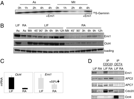
Emi1 levels are high in ES cells. (A) Emi1 inhibits APC/C activity in ES cells. Degradation assay is performed with nonsynchronized or mitotic ES cells for the indicated times. In vitro translated 35S-Geminin is used as a substrate. Recombinant N-terminal–deleted Emi1 is used to inhibit the degradation. Autoradiography for 35S-Securin is shown. (B) Emi1 protein levels are decreased upon differentiation. ES cells are maintained in leukemia inhibitory factor (LIF) containing medium or treated with RA for 48 h to induce differentiation. Cells are synchronized in mitosis and released for the indicated times. Immunoblotting analysis for the indicated proteins is shown. Oct4 is used as a marker for pluripotency. (C) RNA levels for Emi1 are decreased during differentiation. RNA levels for Emi1 and Oct4 are determined by quantitative PCR (qPCR) performed on three independent biological samples synchronized at different time points during S-G2-M progression. Reduction in mRNA for Emi1 is shown. Samples are normalized to β-actin. (D) Emi1 levels associated to APC/C are decreased in differentiated cells. Control cells and cells treated with RA are synchronized in mitosis and immunoprecipitations of Cdc20 or Oct4 (negative control) are performed before immunoblotting analysis for the indicated proteins. Lanes 1 and 2 correspond to 2.5% input.

