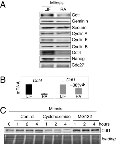
Fig. 4.
Cdt1 is abundant in ES cells. (A) Cdt1 protein levels are higher in mitotic ES cells than in differentiated cells. Immunoblotting for the indicated proteins is shown. Oct4 and Nanog are used as markers of pluripotency. Cdc27 phosphorylation is used as a marker of equal synchronization in mitosis. Mitotic extract of ES cells and differentiated cells (after standard 48-h treatment with RA) are used. (B) RNA levels for Cdt1 are decreased during differentiation. RNA levels for Cdt1 and Oct4 are determined by qPCR performed on three independent biological samples synchronized at different time points during S-G2-M progression. Reduction in mRNA level for Cdt1 is shown. Samples are normalized to β-actin. (C) Cdt1 turnover in mitosis is fast and protein levels are maintained high. Mitotic ES cells are treated with protein-synthesis inhibitor cycloheximide or proteasome inhibitor MG132 for the indicated times. Immunoblotting for Cdt1 protein is shown and equal loading is evaluated by Ponceau staining.

