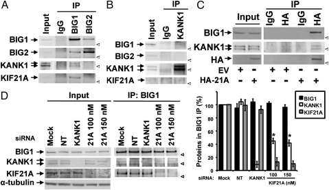
Fig. 1.
Immunoprecipitation of KANK1 with BIG1. (A and B) Samples of Input (2.5% of total) and 25% of proteins from IP with antibodies against BIG1, BIG2, or KANK1, or control IgG from extracts of HeLa cells (1 mg) were separated by SDS/PAGE before reaction of Western blots with indicated antibodies. (A and B are enlarged in Fig. S1.) (C) Samples (50%) of proteins precipitated with anti-HA antibodies or control IgG from 200 μg of extracts prepared 24 h after transfection of cells with HA-KIF21A (HA-21A) or empty vector (EV) were used for Western blotting with indicated antibodies. Input (20 μg) was 10% of amount used for IP. (D) Cells transfected with 100 nM nontargeted (NT) or KANK1-specific siRNA or 100 or 150 nM KIF21A-specific siRNA or with vehicle alone (Mock) were lysed 48 h later. Proteins precipitated with antibodies against BIG1 were analyzed by Western blotting and densitometric quantification. Amounts of proteins from BIG1 IP in three experiments expressed relative to that of the same protein in Mock cells (=100%) are means ± SEM *P < 0.005 vs. Mock. Arrow: protein band. Arrowhead: position of 160-kDa marker.

