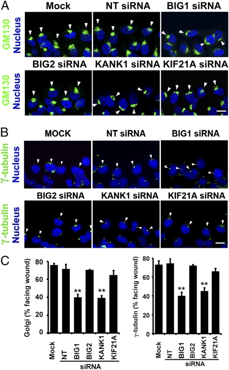
Fig. 4.
Depletion of BIG1 or KANK1 interfered with cell polarization during wound healing. Confluent monolayers of HeLa cells transfected 48 h before with indicated siRNA or vehicle alone (Mock), as in Fig.3, were wounded and fixed 6 h later for staining with DAPI and anti-GM130 (A) or anti-γ-tubulin antibodies (B) and confocal immunofluorescence microscopy. Arrowheads indicate GM130 (A) or γ-tubulin (B). (Scale bar, 10 μm.) (C) Percentage of wound-edge cells with Golgi or γ-tubulin structures in forward-facing 120° sector between nucleus and wound was recorded for at least 100 cells of each population for Golgi localization and 30 cells for γ-tubulin in each experiment. Data are means ± SEM of values from six experiments. **P < 0.02.

