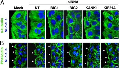
Fig. 5.
Effects of BIG1, BIG2, KANK1, or KIF21A depletion on intracellular microtubule and actin morphology 6 h after wounding. HeLa cells treated as in Figs. 3 and 4 were fixed 6 h after wounding and reacted with anti-α-tubulin antibodies to mark microtubules (A) or Alexa Fluor 488-conjugated phalloidin for F-actin (B). Patterns of microtubules and F-actin were altered in cells depleted of any of the four proteins, but effects on F-actin were most obvious and prominent in cells treated with BIG1 or KANK1 siRNA. (Scale bar, 10 μm.) Arrowheads indicate wound-edge membrane.

