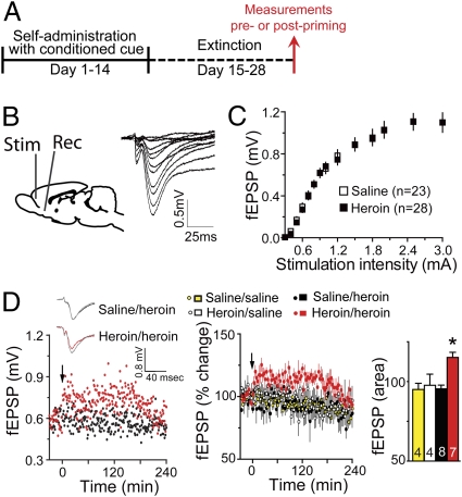
Fig. 1.
A heroin priming injection produces LTP-like changes in fEPSP in the NAcore of heroin-extinguished rats. (A) Heroin treatment protocol used for data in all figures. (B) (Left) Diagram showing the protocol of fEPSP recording in PFc-NAcore excitatory pathway (Rec, recording site; Stim, stimulation site). (Right) Examples of increasing fEPSP amplitude measured in the NAcore of a heroin-extinguished rat in response to increasing stimulation currents in the PFc. (C) Heroin did not alter the input–output relationship between stimulation strength in the cortex and fEPSP amplitude in the NAcore. (D) Acute heroin (0.25 mg/kg, s.c.) induced an increase in fEPSP amplitude for up to 180 min after injection only in heroin-extinguished animals. (Left) Examples of heroin-induced increase in fEPSP amplitude in heroin-extinguished rat (heroin/heroin), but not in saline-yoked control (saline/heroin). (Inset) Sample traces of fEPSP before and after (gray) heroin priming. (Center) Mean ± SEM for all animals. (Right) Area under the curve for 3 h after acute heroin administration. Arrow indicates heroin or saline administration. *P < 0.01, comparing after heroin priming injection in heroin-extinguished animals (heroin/heroin) to heroin in saline-yoked (saline/heroin), saline in heroin-extinguished (heroin/saline), and saline in saline-yoked (saline/saline) using Bonferroni post hoc.

