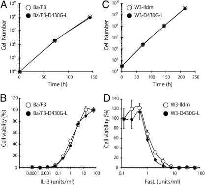
Fig. 2.
No effect of phosphatidylserine exposure on cell growth or cytokine responses. (A) Mouse Ba/F3 and Ba/F3–D430G-L cells were seeded at 104 cells/mL in 1 mL of RPMI1640 medium containing 10% FCS and 100 units/mL mouse IL-3, and cultured at 37 °C for 6 d. The cells were split 1:100 every 3 d, and their growth was followed. (B) Ba/F3 and Ba/F3–D430G-L cells were washed with RPMI1640 containing 10% FCS and cultured at 37 °C for 48 h in medium containing the indicated concentration of mouse IL-3. The cell viability was then determined by the WST-1 assay and is expressed as the percentage of that obtained with 64 units/mL IL-3. (C) Mouse W3-Ildm and W3-D430G-L cells were seeded at 3 × 10⁴ cells/mL in 1 mL of DMEM containing 10% FCS and cultured at 37 °C for 9 d. The cells were split 1:80 every 3 d, and the cell growth was followed. (D) W3-Ildm and W3-D430G-L cells were incubated at 37 °C for 4 h with the indicated concentration of human FasL. The cell viability was then determined by the WST-1 assay and is expressed as the percentage of that without FasL.

