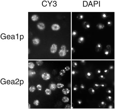
Figure 8.
Gea1p and Gea2p show different localization patterns. Strains YAS61 (Gea2p-myc-his6) and YAS88 (Gea1p-myc) were grown in YPD to early to mid log phase and prepared for immunofluorescence. The GEFs were stained with anti-myc antibodies followed by anti-mouse coupled to CY3 antibodies. The DNA was visualized with 4′,6-diamidino-2-phenylindole.

