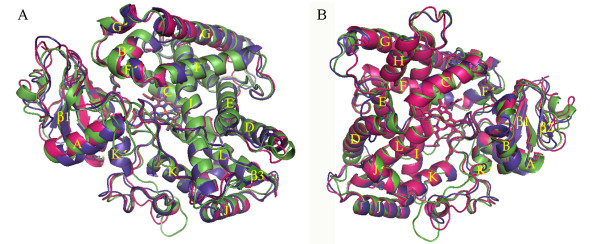
Figure 1.
Overall fold and overlay of homology models of CYP6AA3 (green), CYP6P7 (purple), and CYP6P8 (magenta). Model structures are shown in top (A) and back (B) views. Secondary structures of helices A-L and sheets β1-4 are labeled. The heme group in the middle of the structure is represented by stick.

