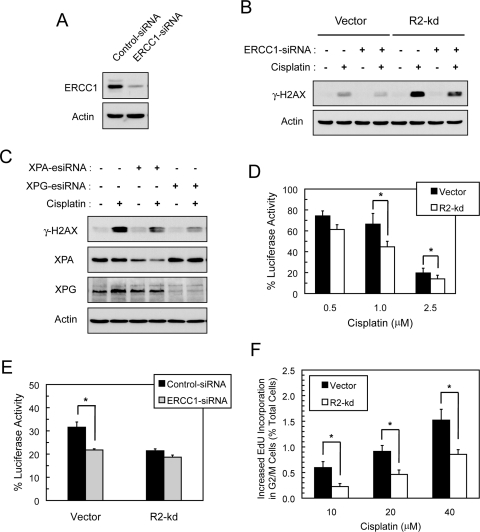
Fig. 6.
R2-knockdown p53(−/−) HCT116 cells exhibited a decrease in NER of cisplatin-induced DNA damage. A, vector cells were transiently transfected with 50 nM control- or ERCC1-siRNA. At 48 h after transfection, cells were harvested and the protein levels of ERCC1 and actin were assessed by Western blotting. B, depletion of ERCC1 attenuated cisplatin-induced γ-H2AX. Vector and R2-knockdown cells were transiently transfected with 50 nM control-siRNA or ERCC1-siRNA. At 24 h after transfection, cells were treated with 10 μM cisplatin for 24 h, and total protein was examined for the levels of γ-H2AX and actin by Western blotting. C, XPA and XPG depletion attenuated cisplatin-induced γ-H2AX in R2-knockdown cells. R2-knockdown cells were transiently transfected with 50 nM control-siRNA, XPA-esiRNA, or XPG-esiRNA. At 24 h after transfection, cells were treated with 10 μM cisplatin for 24 h and total protein was examined for the levels of γ-H2AX and actin by Western blotting. The XPA protein levels after knockdown were 49 and 36% of control for untreated and cisplatin-treated cells, respectively. D, host cell reactivation of cisplatin-damaged luciferase reporter plasmid in p53(−/−) HCT-116 cells. The pGL3 plasmid was treated with the indicated cisplatin concentrations and then transfected into cells. At 48 h after transfection, cells were harvested and assayed for luciferase activities to determine the percentage recovery from cisplatin-induced damage to the plasmid DNA. Values are the means ± S.E. from three independent experiments. *, p < 0.05, paired t test. E, effects of ERCC1 depletion on host cell reactivation of cisplatin-damaged luciferase reporter plasmid in p53(−/−) HCT-116 cells. Cells were cotransfected with control- or ERCC1-siRNA and pGL3 plasmid treated with 1 μM cisplatin, and then harvested at 48 h after transfection. Luciferase activities were measured and calculated as described in D. Values are means ± S.E. from a representative experiment of two, each performed in triplicate. *, p < 0.05, paired t test. F, R2-knockdown cells displayed a decrease in cisplatin-induced EdU incorporation at the G2/M phase of the cell cycle. Cells were treated with 0, 10, 20, and 40 μM cisplatin together with 10 μM EdU for 4 h. Subsequently cells were fixed, stained, and analyzed by flow cytometry. Bivariate distribution of EdU incorporation and DNA content was used to determine increases in cisplatin-induced EdU incorporation in the G2/M cell population above the values of untreated controls. Values are percentages of total cells sorted and expressed as the means ± S.E. from three independent experiments. *, p < 0.05, paired t test.

