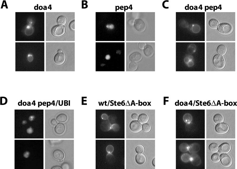
Figure 4.
Intracellular localization of Ste6-GFP fusions. GFP fluorescence was observed in exponentially growing cells transformed with pRK599 (wild-type STE6 fused to GFP) or pRK628 (STE6 ΔA-box fused to GFP). (A) JD116/pRK599 (doa4). (B) RKY975/pRK599 (pep4). (C) RKY1449/pRK599 (doa4 pep4). (D) RKY1449/pRK599/YEp96 (doa4 pep4, 2μ-ubiquitin). (E) JD52/pRK628 (wild type). (F) JD116/pRK628 (doa4). Left, GFP-fluorescence; right, differential interference contrast (DIC) image. The cells were grown at 30°C in minimal medium with 1% casamino acids.

