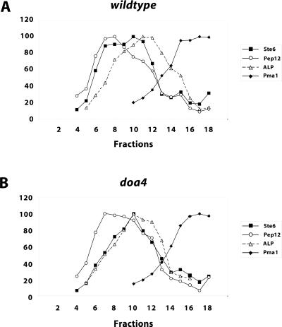
Figure 6.
Distribution of marker proteins from wild-type and doa4 strains on sucrose gradients. Whole cell extracts of the wild-type strain JD52 (A) and the doa4 strain JD116 (B) grown at 30°C in minimal medium with 1% casamino acids were fractionated on a sucrose density gradients (20–50% [wt/wt] sucrose, fraction 1: low sucrose density). The gradient fractions were analyzed by Western blotting for the presence of marker proteins with specific antibodies. The Western blots were scanned and the signal intensities were quantified with the program NIH Image 1.61. The strongest signals were set to 100%. ▪, Ste6; ○, Pep12; ▵, ALP; ♦, Pma1.

