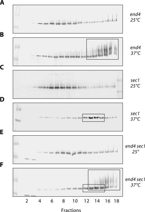
Figure 7.
Sec1-independent Ste6 transport to the plasma membrane. The intracellular distribution of Ste6 in different strains, transformed with the single-copy STE6 plasmid pRK182, was examined by sucrose density gradient fractionation (20–50% [wt/wt] sucrose, fraction 1: low sucrose density) and Western blotting with anti-Ste6 antibodies. The strains were either continuously grown at 25°C (A, C, and E) or were first grown at 25°C and then shifted to 37°C for 1 h (B, D, and F) before extract preparation. (A and B) RKY1203/pRK182 (end4). (C and D) RKY1190/pRK182 (sec1-1). (E and F) RKY1177/pRK182 (end4 sec1-1). The area where the plasma membrane marker Pma1 is found in the gradients is marked by large boxes in B and F. The putative secretory vesicle peak is highlighted by small boxes in D and F.

